next up previous contents
Next: 5.4 Changes in the Up: 5 Aftereffect Experiments Previous: 5.2 Angular function of

5.3 Changes in the patterns of connection strength

  The TAE magnitudes shown in figure 5.3 result from changes in the response to the test line after adaptation. Each point on the graph represents the change in the overall activity distribution for a test line, so it is an aggregate measure of many small changes. To illustrate the changes in more detail, this section examines how the individual weights change with adaptation. For clarity, only inhibitory weights were allowed to be modified ($\alpha_I=0.00005$ and $\alpha_A=\alpha_E=0$). Figure 5.4 shows how the connections of a typical neuron in the central region of the model cortex changed during adaptation.


Figure 5.4: Weights of a central neuron before and after adaptation.
 These plots show the strength of the weights of each type going to the neuron at (91,98), which is marked with a tiny box in the cortex plots of (c) and (d). The afferent weights (a) of this neuron have an orientation preference of +45°, and were not modifiable in this experiment. The fixed excitatory weights in (b) connect only to the nearest neighbors in the cortex. Those neurons all have similar orientation preferences, so even if these weights had been modifiable they would not have changed the response of the neuron significantly. The inhibitory connections span a wide range of locations and orientation preferences. They undergo large and orientation-specific changes with adaptation. Comparing ( c) with (d) shows that the blue areas along the vertical line down the center have increased in strength as a result of adapting to the vertical test line.
\begin{figure}
\begin{minipage}
{\textwidth}
 \centering
 \begin{minipage}
{\two...
 ...\baselineskip}\newline
\usebox {\orkey}

 \end{minipage} \newline

 \end{figure}

Adaptation has redistributed the inhibitory weights. Connections to neurons that have orientation preferences similar to the adaptation line became stronger (the blue areas in figure 5.4d). The strengthening results in the direct effect: inhibition for orientations close to the adaptation orientation is increased. However, since total weight strength for all inhibitory weights is constant (equation 3.4), the connections to other orientations (the yellow and red areas) must simultaneously decrease. This causes the indirect effect: inhibition for distant orientations is reduced. Chapter 6 discusses possible biological mechanisms for this weight normalization.

Since the changes for each neuron are relatively small, the difference may not be readily apparent from figure 5.4. Some changes are visible on close inspection in the central blue and red-orange areas; compare figure 5.4 ( c) and (d). To highlight the changes, figure 5.5 shows the result of subtracting the weight plots from before and after adaptation.
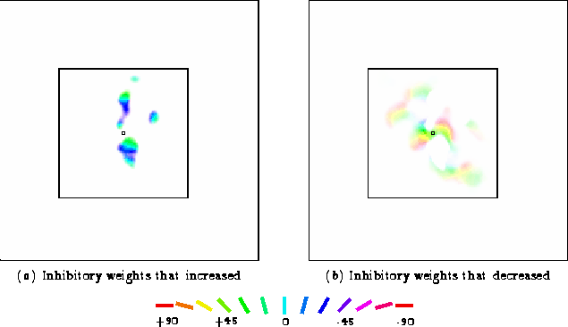
 
Figure 5.5: Weight changes for a central neuron with adaptation.
  These plots show the weight changes for the neuron in figure 5.4 in more detail. Colored areas in (a) represent inhibitory connections that increased in strength. The plot was computed by subtracting the weights in figure 5.4c from those in figure 5.4d, scaling the value up to a visible level, then labeling each connected neuron with the color corresponding to its orientation preference. Only connections within the cortical area corresponding to the input Gaussian have been strengthened, and only those whose orientation preferences are close to the adaptation angle (i.e., neurons colored blue, cyan, or green). These changes cause the direct tilt aftereffect, as explained in section 5.4.2. While those connections strengthened, the rest of the connections to that neuron had to weaken (due to equation 3.4). The colored areas in (b) represent inhibitory connections that decreased in strength. The plot was computed by subtracting figure 5.4d from figure 5.4c, scaling it by the same factor as in (a), and labeling each point with the orientation preference of the connected neuron. The weakened connections are all those present for this neuron other than those that were strengthened; each was weakened in proportion to its current strength. These changes cause the indirect tilt aftereffect, as explained in section 5.4.3.
\begin{figure}
\begin{minipage}
{\textwidth}
 \centering
 \begin{minipage}
{\two...
 ...\baselineskip}\newline
\usebox {\orkey}

 \end{minipage} \newline

 \end{figure}

Subtracting the before plot from the after plot shows which connections have increased in strength, and subtracting the after plot from the before plot shows which ones have decreased. Clearly, inhibitory connections to orientations near the adaptation line have increased in strength, while those to distant orientations have decreased. These systematic changes in the inhibitory weights result in both direct and indirect tilt after-effects, as seen in the next section.


next up previous contents
Next: 5.4 Changes in the Up: 5 Aftereffect Experiments Previous: 5.2 Angular function of
James A. Bednar
9/19/1997